
Echocardiography
Abstract
Echocardiography is one of the most relevant non-invasive diagnostic tools for real-time imaging of cardiac structure and function. Significant advances in 3-D transthoracic echocardiography (TTE) and 3-D transesophageal echocardiography (TEE) have made this modality a powerful tool in the clinic. Real-time 3-D TEE, for instance, has become the standard echocardiographic modality for visualization of structures in the atrial and valvular regions of the heart. Furthermore, ultrasound (US) data are generated by reflection of transmitted coherent ultrasound waves at fixed frequencies. The result of the interaction between those waves and the different types of tissues give rise to interference phenomenon known as speckle. This interference pattern, albeit its textured and noisy visual aspect, remains unaltered under the same acquisition conditions, i.e., the same transducer aperture, pulse length and transducer angle. This behavior suggests that speckle exhibits an inherent relationship with the tissue structure that can be used to estimate the motion of heart
Strain and Motion Estimation - Speckle Tracking
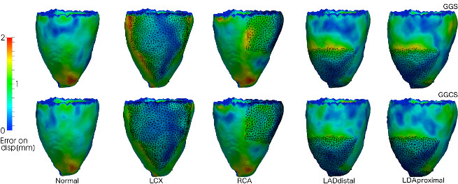
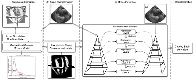
In echocardiography (EC), the strain analysis became possible using Tissue Doppler Imaging (TDI). Unfortunately, this modality shows an important limitation: the angle between the myocardial movement and the ultrasound beam should be small to provide reliable measures. This constraint makes it difficult to provide strain measures of the entire myocardium. Alternative non-Doppler techniques such as Speckle Tracking (ST) can provide strain measures without angle constraints. However, the spatial resolution and noisy appearance of speckle still make the strain estimation a challenging task in EC. Several maximum likelihood approaches have been proposed to statistically characterize the behavior of speckle, which results in a better performance of speckle tracking. However, those models do not consider common transformations to achieve the final B-mode image (e.g. interpolation). We work on different maximum likelihood approaches for speckle tracking which effectively characterizes speckle of the final B-mode image. Moreover, the Speckle tracking accuracy is improved by using probabilistic tissue characterizations. A typical approach for strain and motion estimation can be summarized in next figure:
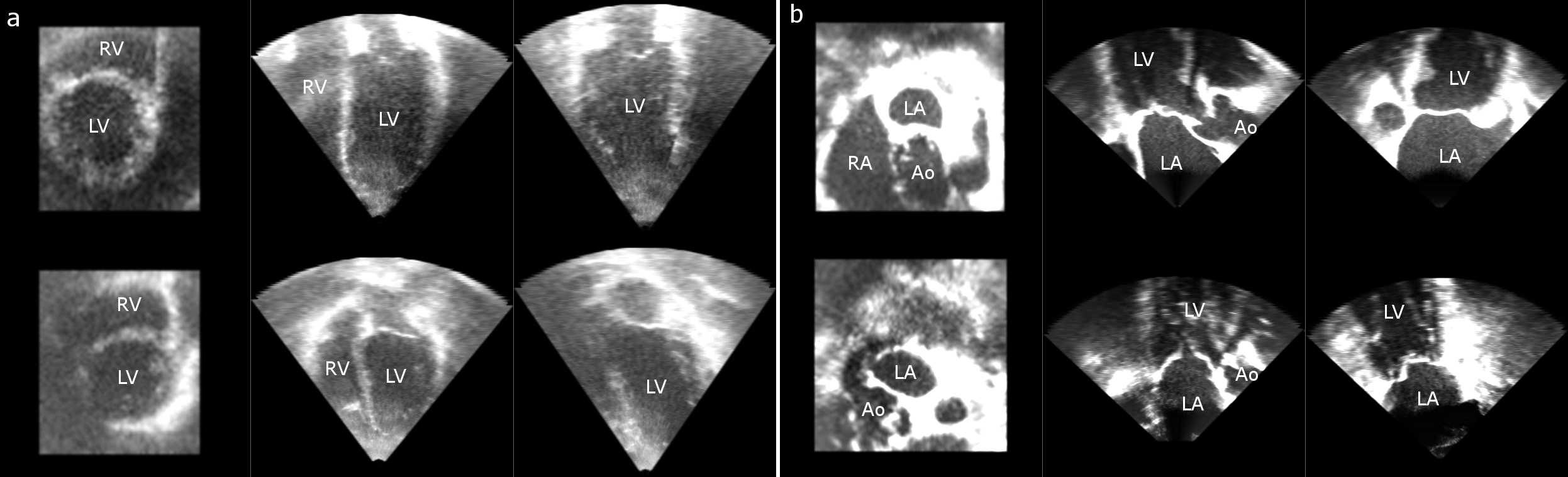
Cardiac Structure Detection
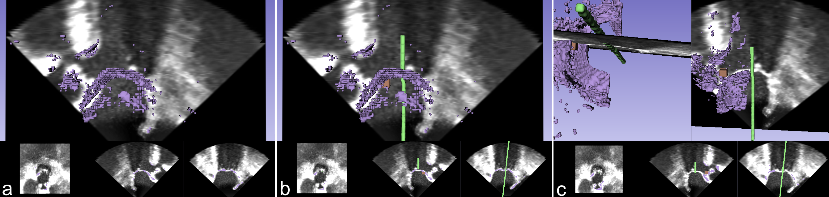
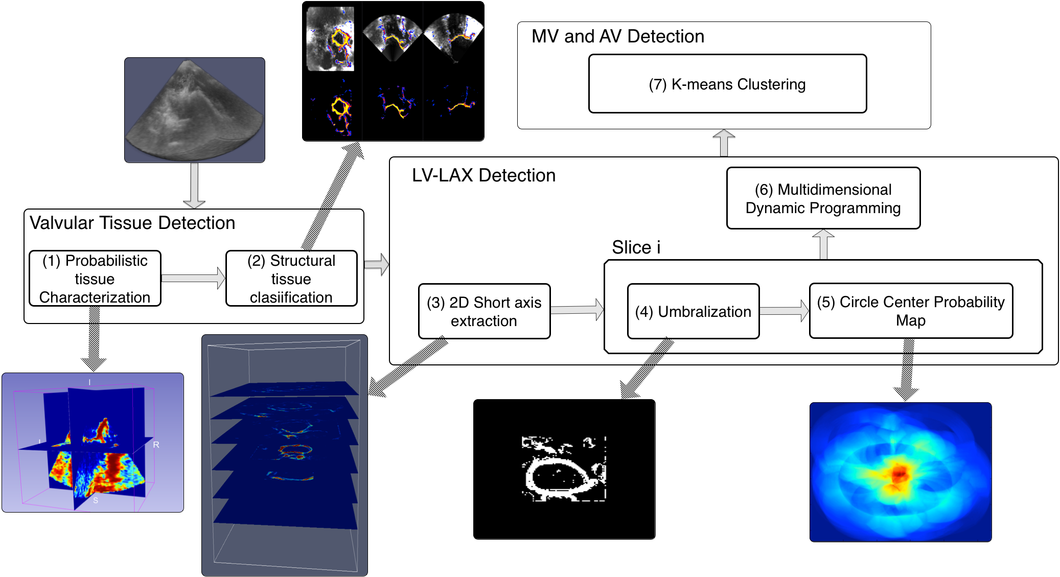
Most automated segmentation approaches to the mitral valve and left ventricle in 3-D echocardiography require a manual initialization. We work on a fully automatic scheme to initialize a multicavity segmentation approach in 3-D transesophageal echocardiography by detecting the left ventricle long axis, the mitral valve and the aortic valve location. Our approach uses a probabilistic and structural tissue classification to find structures such as the mitral and aortic valves; the Hough transform for circles to find the center of the left ventricle; and multidimensional dynamic programming to find the best position for the left ventricle long axis. The workflow for the methodology used to detect the left ventricle longaxis (LV-LAX), mitral valve(MV) and aortic valve (AV) is summarized in next figure:
Papers
- A. H. Curiale, G. Vegas-Sánchez-Ferrero, J. G. Bosch and S. Aja-Fernández, “A Maximum Likelihood Approach to Diffeomorphic Speckle Tracking for 3D Strain Estimation in Echocardiography,” Medical Image Analysis, Available online 23 May 2015, DOI: 10.1016/j.media.2015.05.001
- A. H. Curiale, G. Vegas-Sánchez-Ferrero and S. Aja-Fernández, “Probabilistic Tissue Characterization for Ultrasound Images,” Insight Journal (2015), http://hdl.handle.net/10380/3517.
- A. H. Curiale, A. Haak, G. Vegas-Sánchez-Ferrero, B. Ren, S. Aja-Fernández, and J. G. Bosch, “Fully automatic detection of salient features in 3-d transesophageal images,” Ultrasound in Medicine & Biology, vol. 40, no. 12, pp. 2868–2884, 2014.
- A. H. Curiale, G. Vegas-Sánchez-Ferrero, and S. Aja-Fernández, “Speckle tracking in interpolated echocardiography to estimate heart motion,” in Functional Imaging and Modeling of the Heart (S. Ourselin, D. Rueckert, and N. Smith, eds.), vol. 7945 of Lecture Notes in Computer Science, pp. 325– 333, Springer Berlin Heidelberg, 2013.
- A. H. Curiale, G. Vegas-Sanchez-Ferrero, T. Perez-Sanz, and S. Aja-Fernandez, “Strain rate tensor estimation from echocardiography for quantitative assessment of functional mitral regurgitation,” in Biomedical Imaging (ISBI), 2013 IEEE 10th International Symposium on, pp. 788–791, 2013.
- A. H. Curiale, G. Vegas-Sánchez-Ferrero, T. Pérez-Sanz, and S. Aja-Fernández, “Cuantificación de la insuficiencia mitral funcional mediante el esfuerzo y la velocidad del miocardio,” in XXIX Congreso Anual de la Sociedad Española de Ingeniería Biomédica, pp. 321–324, Centro de Cirug ́ıa de Mínima Invasión Jesús Usón, 2011.